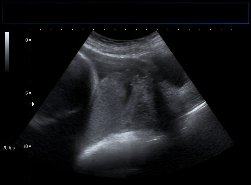
Muskuloskeletal / 3d/4d sonography
Home / Muskuloskeletal / 3d/4d sonography
All Services

Overview
Using high-frequency sound waves, musculoskeletal sonography, also referred to as musculoskeletal ultrasound, allows real-time visualization of soft tissues such as joints, tendons, ligaments, and muscles. This imaging modality is very helpful in guiding injections, evaluating soft tissue and joint problems, and diagnosing and tracking musculoskeletal injuries such as rips, strains, and inflammation.
3D and 4D sonography build upon traditional 2D ultrasound by providing more detailed and dynamic views:
3D Sonography creates three-dimensional images, which can offer a more detailed view of the anatomy compared to 2D images. This is useful for assessing complex structures and abnormalities in greater detail.
4D Sonography adds a time element to the 3D images, allowing for real-time motion visualization. This technique can be particularly beneficial for observing dynamic movements, such as joint function or fetal development in obstetrics.
Both 3D and 4D sonography are valuable in fields like orthopedics and sports medicine for providing comprehensive assessments of musculoskeletal conditions and facilitating more accurate diagnoses and treatment plans.
