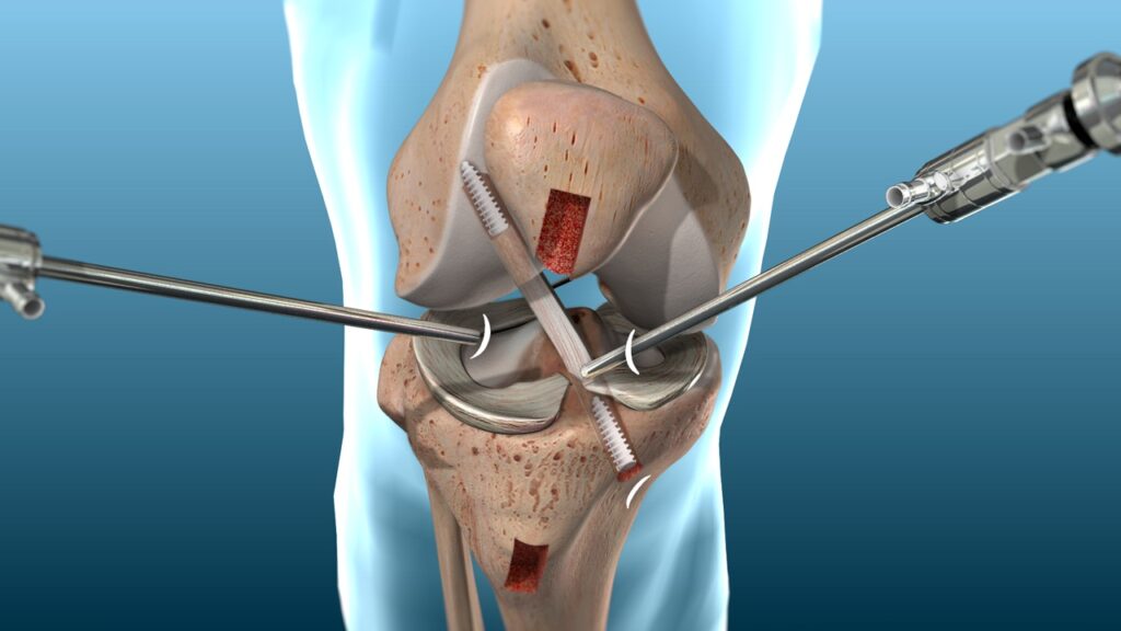
ACL Surgery
Home / ACL Surgery
All Services

ACL Surgery: How Diagnosis and Imaging Tests Help Assess Knee Injuries
In order to compare your injured and uninjured knees, your doctor will examine your knee during the physical examination to look for swelling and discomfort. In order to evaluate the joint’s general function and range of motion, he or she may additionally move your knee in a number of ways.
Often the diagnosis can be made on the basis of the physical exam alone, but you may need tests to rule out other causes and to determine the severity of the injury. These tests may include:
- X-rays. X-rays may be needed to rule out a bone fracture. However, X-rays don’t show soft tissues, such as ligaments and tendons.
- Magnetic resonance imaging (MRI). An MRI uses radio waves and a strong magnetic field to create images of both hard and soft tissues in your body. An MRI can show the extent of an ACL injury and signs of damage to other tissues in the knee, including the cartilage.
- Ultrasound. Using sound waves to visualize internal structures, ultrasound may be used to check for injuries in the ligaments, tendons and muscles of the knee.
